Loading 3D models...
COVID Spike Protein
by wgurley in science-technology
644,446
Faces
326,355
Vertices
0
Textures
Free
Download
Description
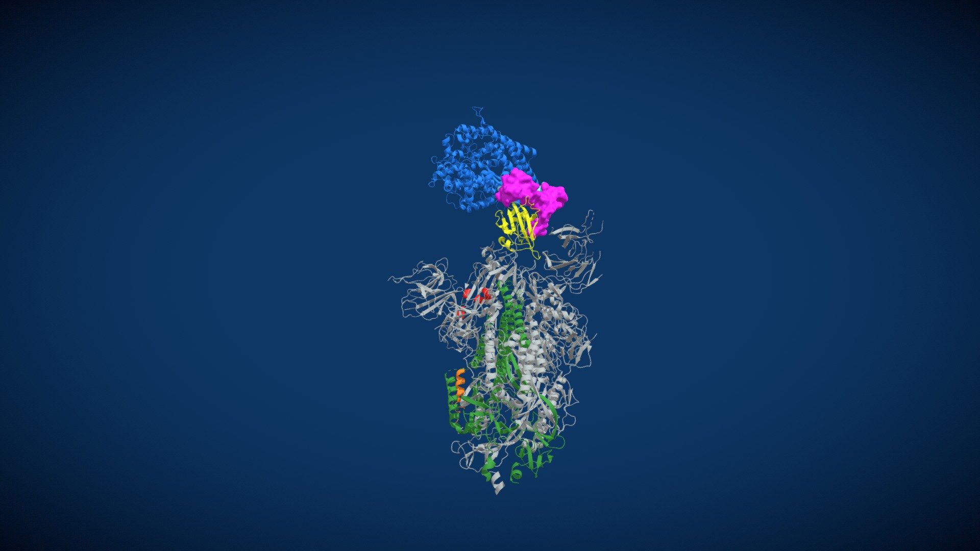
Molecular image from the RCSB Protein Data Bank, file 6VSB. **Pale yellow** = S1 domain (attaches to the ACE2 target cell receptor). **Green** = Stem domain (S2), which unfolds upon activation to embed transmembrane regions into the membrane of the target cell. **Red** = domain that is removed by two cuts from the target cell protease (membrane protease). **Magenta** = site of the first cut, which releases the S1 domain, which is attached to the ACE2 receptor. **Organge-yellow** = (between red & green regions) site of the second cut, which activates the hinged fusion protein extended conformation. **Blue** = Fusion protein domain (embeds into the target cell membrane).
Technical Details
Related Models
EdgeSword - Sword
12,770 faces
Free
COVID-19 test line in Aarhus
0
Free
Barrette Arch
71,026 faces
Free
nucleocapsid-RNA complex of rhabdovirus
2,633,848 faces
Free

Covid Bin
13,090 faces
Free
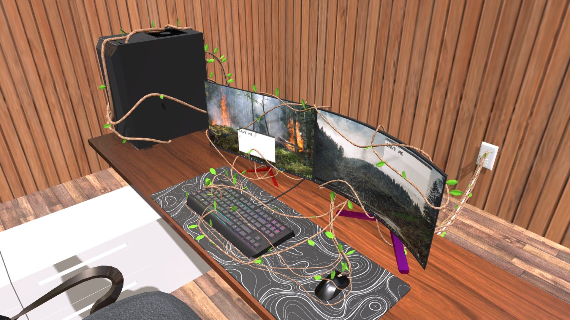
Mother nature collides with Digital era
441,321 faces
Free
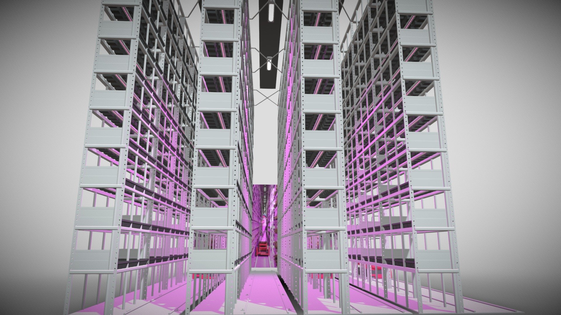
GROWING PLANTS
2,404,560 faces
Free
7 Spike-s2h97 Site
347,076 faces
Free